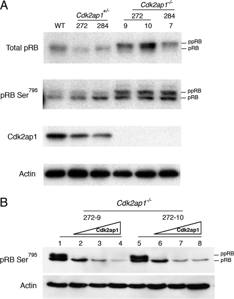
FIGURE 3.
Hyperphosphorylation of pRb in Cdk2ap1−/− mESCs. A, the level of pRb and phosphorylation status of serine 795 of pRb was examined by Western analysis by using specific antibodies (anti-pRb G3–245 from BD Pharmingen and anti-pRb Ser795 antibody from Cell Signaling Technology, Inc. (Danvers, MA)). Cdk2ap1−/− cells (clones 272-9, 272-10, and 284-7) showed induced hyperphosphorylation of pRb compared with WT or their parental Cdk2ap1+/− clones (272 and 284). B, the effect of restoring Cdk2ap1 on pRb phosphorylation was examined in Cdk2ap1−/− mESCs. Cells were transduced with control virus (lanes 1 and 5) or Ad-CDK2AP1 virus (lanes 2 and 6, MOI 10; lanes 3 and 7, MOI 20; lanes 4 and 8, MOI 30) as shown in Fig. 1. The result showed hypophosphorylation of pRb upon the expression of CDK2AP1, demonstrating the specificity of the role of CDK2AP1 in pRb phosphorylation.

