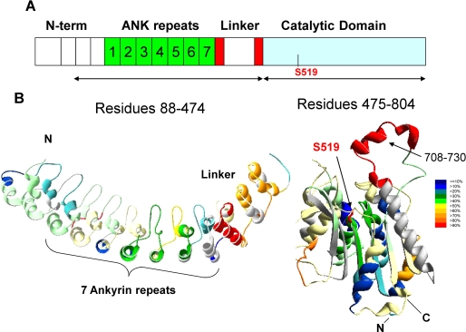
FIGURE 4.
Deuteration level of GVIA-2 iPLA2. A, schematic representation of GVIA-2 iPLA2. The seven ankyrin repeats are in green. Three helix-turn-helix-loop structures are the three blocks upstream of ankyrin repeats. The eighth ankyrin repeat (red) is interrupted by a 54-residue insertion (white). B, the deuterium exchange map of GVIA-2 iPLA2 is shown after 3000 s of on-exchange with the color coding indicating the percentage of hydrogen/deuterium exchange.

