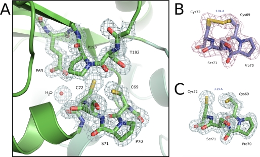
FIGURE 3.
Active site region of BdbD. A, detailed view of the N terminus of helix α1 of sBdbD showing the Cys-Pro-Ser-Cys active site of sBdbD and the closely lying cis-proline (Pro193), which is invariant in all thioredoxin-like proteins. B and C, electron density (contoured at 1.0σ) of the active site region of sBdbD in oxidized and reduced states, respectively.

