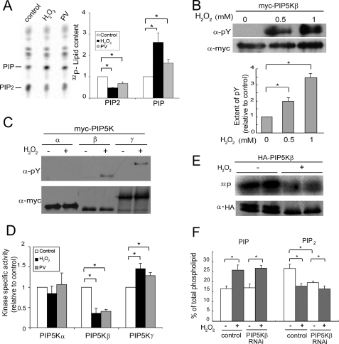
FIGURE 2.
H2O2 induces PIP5Kβ tyrosine phosphorylation and decreases its lipid kinase activity. A, PV and H2O2 have similar effects on phosphoinositide homeostasis. HeLa cells labeled with [32P]orthophosphate were treated with either 1 mm H2O2 or 10 μm PV for 15 min. Lipids were extracted and resolved by TLC. Left, fluorogram; right, quantitation. The amount of PIP2 or PIP in the control sample was set as 1. B, PIP5Kβ tyrosine phosphorylation. COS cells transfected with Myc-PIP5Kβ were exposed to 0.5 or 1 mm H2O2 for 20 min. Myc-PIP5Kβ was immunoprecipitated and Western blotted with α-Myc and α-Tyr(P). Top, Western blots; bottom, quantitation of tyrosine phosphorylation. The ratio of α-Tyr(P) to α-Myc PIP5Kβ intensity in the absence of H2O2 is set as 1. Values shown are mean ± S.E., n = 3. Asterisks denote statistically significant compared with control, with p < 0.05, in this and all other panels in this figure. C, H2O2 induces tyrosine phosphorylation of PIP5Kβ and γ87, but not PIP5Kα. Cells were incubated with 1 mm H2O2 for 15 min. D, H2O2 has differential effects on the lipid kinase activity of the three PIP5K isoforms. COS cells transiently transfected with Myc-PIP5K isoforms were exposed to either 1 mm H2O2 or 10 μm PV for 15 min. Immunoprecipitated Myc-PIP5Ks were used in an in vitro lipid kinase assay and their activity in the linear range was normalized against the amount of immunoprecipitated kinase. The specific activity (mean ± S.E., n = 3) of each control untreated PIP5K isoform was set as 1. E, H2O2 also induces PIP5Kβ Ser/Thr dephosphorylation. COS cells expressing HA-PIP5Kβ were labeled with 32P and exposed to 1 mm H2O2. Immunoprecipitated HA-PIP5Kβ was subjected to SDS-PAGE. 32P-Labeled proteins were detected by PhosphorImager and total HA-PIP5Kβ was detected by Western blotting. Data shown are representative of those from two independent experiments. F, effects of PIP5Kβ depletion by RNAi on the H2O2-induced decrease in PIP2. HeLa cells were transfected with small interfering RNA oligonucleotides targeting PIP5Kβ or firefly luciferase (negative control). Cells were stimulated with H2O2 (0.5 mm, 15 min), and 32P incorporation into PIP and PIP2 was quantitated after TLC. The amount of [32P]phosphoinositide was expressed as a percentage of total labeled phospholipids. Data shown are mean ± S.E., n = 7 from three independent experiments. Error bars indicate S.E.M.

