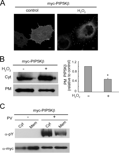
FIGURE 3.
H2O2 decreases PIP5Kβ membrane association. COS cells overexpressing Myc-PIP5Kβ were exposed to 1 mm H2O2 or 10 μm PV for 15 min. A, immunofluorescence localization. Cells were stained with α-Myc/FITC. The periphery of the cell, based in cortical phalloidin actin staining (not shown), is outlined by arrowheads. Scale bars, 10 μm. B, decrease in PIP5Kβ association with a PM-enriched fraction. Cell homogenates were subjected to sequential multistep centrifugation. The PM-enriched and cytosolic (Cyt) fractions were blotted with α-Myc. Left, Western blot; right, change in PM-associated Myc-PIP5Kβ. The ratios of PM/PM + Cyt Myc-PIP5Kβ were plotted (mean ± S.E., n = 3), relative to that of the untreated control set as 1. Asterisk denotes statistically significant, with p < 0.05. C, preferential decrease in tyrosine-phosphorylated Myc-PIP5Kβ from microsome membranes. Cytosol (Cyt) and microsome membranes (Mem) from PV-treated cells were separated and blotted with α-Tyr(P) and α-Myc. The Tyr(P)/Myc ratios are 3.54 and 1.86 for Cyt and Mem from H2O2-treated cells. Data shown are representative of two independent experiments. Error bars indicate S.E.M.

