FIGURE 1.
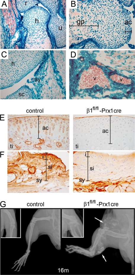
Deletion of the β1 integrin gene in mice using the Prx1-cre transgenic line. A, X-gal staining of a forelimb section from β1fl/fl-Prx1cre+ embryonic day 18.5 embryo shows Cre recombinase activity in cartilaginous and bony tissues of the elbow and in muscle fibroblasts (h, humerus; r, radius; u, ulna). B, distal epiphysis of the humerus exhibits Cre activity in all chondrocytes (blue) of the presumptive articular cartilage (ac), whereas in the growth plate (gp), some chondrocytes (red) are negative. C and D, X-gal staining demonstrates Cre activity in the forming synovium (sy) (C) and in the vessels (v) (D). E and F, β1 integrin immunostaining at 1 month of age demonstrates the absence of β1 integrin on articular cartilage chondrocytes of mutant tibia (ti) (E) and on synoviocytes of the synovium (sy) (F). The bars on F indicate the intimal (i) and subintimal (si) layers of the synovium. G, x-ray of the hind limb at 16 months indicates short bones, joint space narrowing, and blood vessel calcification (arrows) in mutants.

