FIGURE 9.
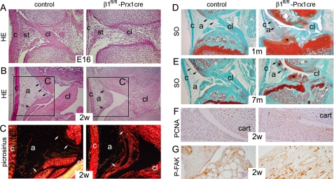
Synovial abnormalities in β1fl/fl-Prx1cre+ mice. A and B, photomicrographs of HE-stained midsagittal sections of the knee joints. A, looser appearance of the developing synovial tissue (st) and the delayed formation of the cruciate ligament (cl) were observed in 16-day-old mutant embryo (E16). Also note the thinner collateral ligament (c) in mutant. B, at 2 weeks (2w), the control synovium consists of a thin lining layer (arrows) and a large adipose (a) tissue. In mutant, the lining is hyperplastic with large vessels, and the amount of adipose tissue is reduced. C, picrosirius staining followed by polarization microscopy of the boxed areas in B reveals fibrosis of the mutant synovium, as shown by the strongly birefringent collagen deposits (white arrows). Also note the reduced birefringence in the mutant cruciate ligament, demonstrating structurally abnormal, less oriented, collagen fibrils in this tissue. D and E, photomicrographs of safranin orange-Fast green (SO)-stained sections. D, at 1 month, the intercondylar tibial surface is cartilaginous in the mutant and covered by the synovial tissue, showing signs of chondrification (asterisk). E, by 7 months, extensive chondrification (asterisks) is observed in the joint space and in the synovial lining (arrows) of mutant. F, increased numbers of PCNA-positive synovial cells are visible in the mutant. G, increased phosphorylation of FAK was evident in fibroblast-like cells of the mutant synovium. All stains were performed on paraffin sections, and the results are representative of those seen in at least four mice of each genotype at each age.

