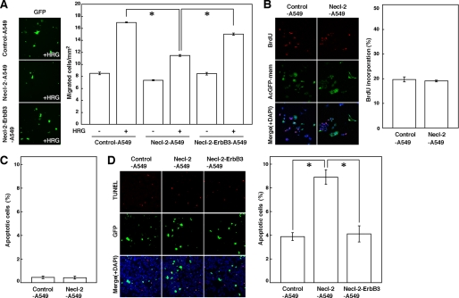
FIGURE 4.
Inhibition by Necl-2 of movement and anchorage-independent survival of A549 cells. A, measurement of cell movement using a Boyden chamber assay. A549 cells were co-transfected with empty vector and pmaxGFP (Control-A549 cells), FLAG-Necl-2 expression vector and pmaxGFP (Necl-2-A549 cells), or FLAG-Necl-2, ErbB3 expression vector, and pmaxGFP (Necl-2-ErbB3-A549 cells), and were plated on cell culture inserts (8.0-μm pore membrane filters) in the presence or absence of 100 ng/ml HRG in the bottom well for 18 h. The EGFP-positive migrated cells were counted by microscopic examination. Error bars indicate the means ± S.E. of three independent experiments. *, p < 0.001. B, measurement of cell proliferation. A549 cells which were co-transfected either with empty vector and pAcGFP-mem or with FLAG-Necl-2 expression vector and pAcGFP-mem (Control-A549 and Necl-2-A549 cells, respectively) were starved of serum and stimulated by HRG in the presence of BrdUrd. After 14 h of incubation, cells were fixed, and then double stained with the anti-BrdUrd mAb and 4′,6-diamidino-2-phenylindole (DAPI). AcGFP-mem-positive cells were measured. The left panels show representative fields of three independent experiments. Error bars indicate the means ± S.E. of three independent experiments. C, measurement of anchorage-dependent apoptotic cells. A549 cells which were co-transfected either with empty vector and pmaxGFP or with FLAG-Necl-2 expression vector and pmaxGFP (Control-A549 and Necl-2-A549 cells, respectively) were starved of serum for 48 h and subjected to a TUNEL assay. Error bars indicate the means ± S.E. of three independent experiments. D, measurement of anchorage-independent apoptotic cells. A549 cells that were co-transfected with empty vector and pmaxGFP (Control-A549 cells), FLAG-Necl-2 expression vector and pmaxGFP (Necl-2-A549 cells), or FLAG-Necl-2, ErbB3 expression vector, and pmaxGFP (Necl-2-ErbB3-A549 cells) were cultured in suspension in the presence of 0.5% fatty acid-free BSA and 10 ng/ml HRG. After 48 h of incubation, the cells were subjected to a TUNEL assay. Error bars indicate the means ± S.E. of three independent experiments. *, p < 0.02.

