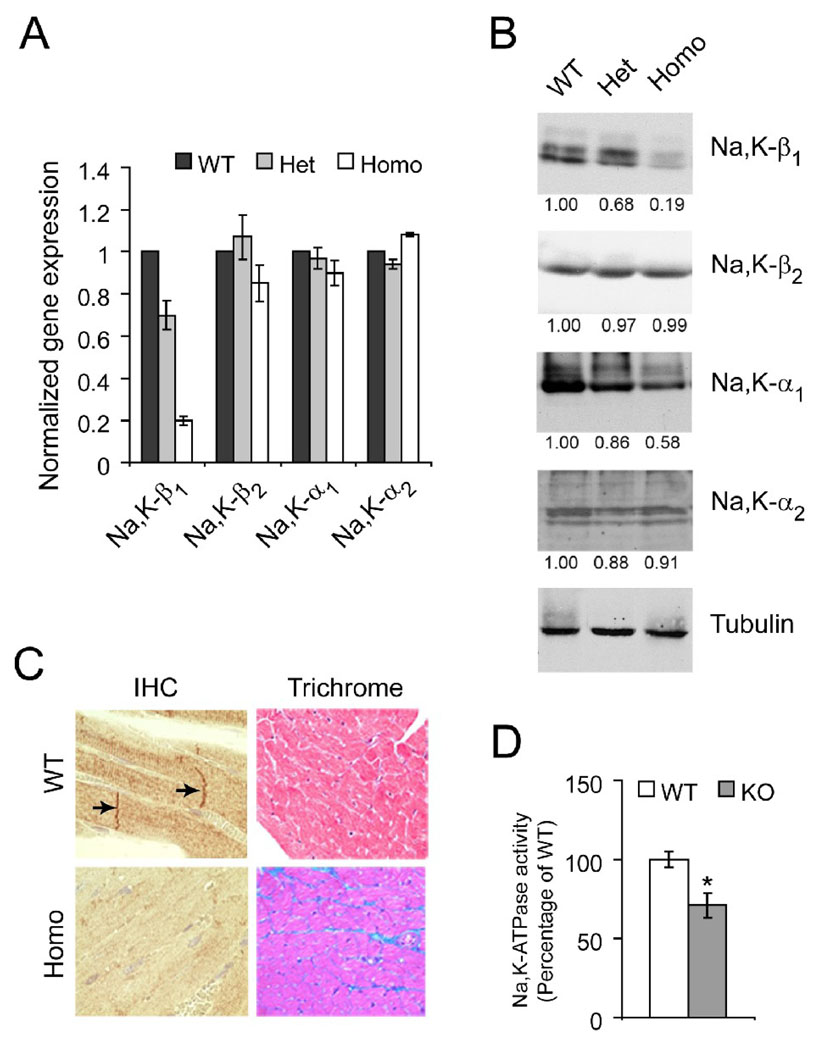
Figure 2.
Characterization of cardiac-specific Na,K-β1 knockout mice. Quantitative RT-PCR (A) and representative western blots (B) from heart lysates of wild-type (WT), heterozygous (Het), and homozygous KO (Homo) mice (n=4 each). The staining of Na,K-β1 as a narrow doublet has also been observed by other investigators in heart lysates. For immunoblots, the densitometric quantitation of the band intensities normalized to the loading control, tubulin are placed below each panel. (C) Comparison of the heart tissues of WT and KO mice by immunohistochemistry (IHC) and trichrome staining. Arrows represent staining of Na,K-β1 in the intercalated discs. Representative images from at least 5 mice are shown. (D) Na,K-ATPase pump activity measured as the amount of ouabain-sensitive inorganic phosphate from sarcolemmal membrane preparations from WT and KO hearts. Values are represented as Mean ± SE (n=3 each, * p < 0.05) and plotted as a percentage of the WT.

