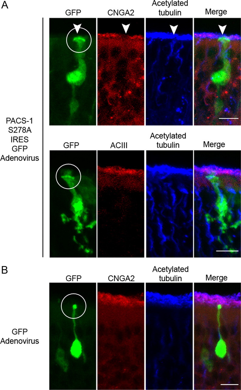
Figure 7.

Expression of mutant PACS-1 in native OSNs causes mislocalization of the CNG channel, but not ACIII. Representative collapsed confocal images of coronal sections of OE from adenovirally infected mice. A, Top, Immunostaining of a PACS-1 S278A IRES GFP-infected OSN (green) with antibodies against CNGA2 (red) and acetylated α tubulin (blue) demonstrates a loss of ciliary CNG channel from the infected OSN (white arrowhead) with no change in the cilia layer. Bottom, Immunostaining of a PACS-1 S278A IRES GFP-infected OSN (green) with antibodies against ACIII (red) and acetylated α tubulin (blue) demonstrates no detectable change in ciliary ACIII or the cilia layer from the infected OSN. B, Immunostaining of a GFP-infected OSN (green) with antibodies against CNGA2 (red) and acetylated α tubulin (blue) demonstrates no detectable change in ciliary CNG channel or the cilia layer from the infected OSN. For all conditions, a merged image is shown on the right (Merge), white circles mark dendritic knobs, and scale bars represent 10 μm.
