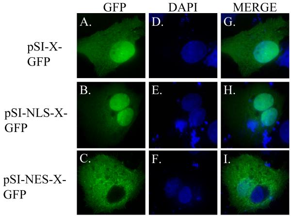
FIG. 4. Subcellular localization of HBx-GFP fusion proteins in HepG2 cells.
HepG2 cells were transfected with pSI-X-GFP, pSI-NLS-X-GFP, or pSI-NES-X-GFP and analyzed by deconvolution microscopy 24 hours post-transfection. Localization of GFP tagged constructs is illustrated in the first column (A-C), staining of the cell nucleus (by DAPI) can be seen in the middle column (D-F), and a merged image can be seen in the right column (G-I).

