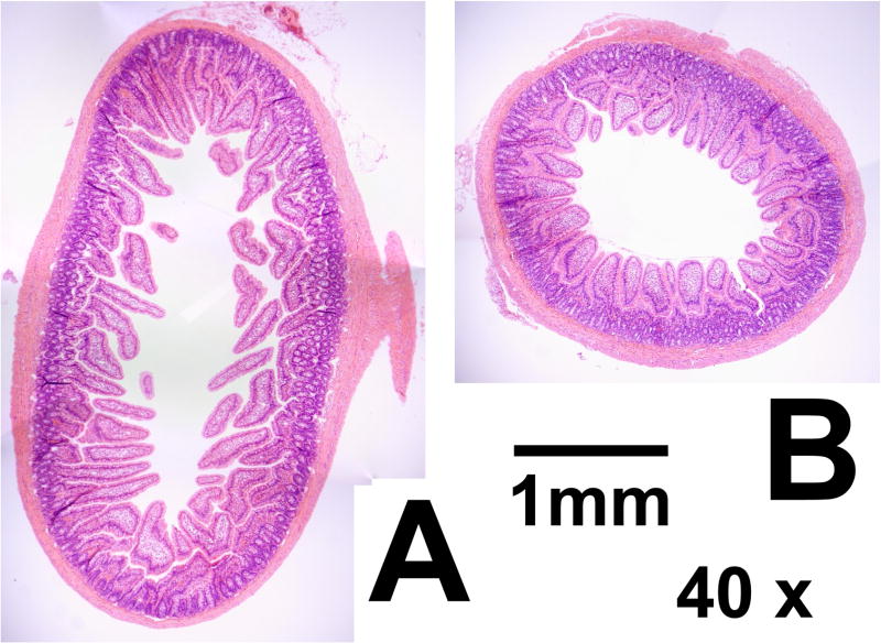
Figure 2.
Representative composite photomicrographs of sections from JEJ (A) and LOOP (B) under 40x magnification. Both sections are from the same animal. The increased diameter in JEJ sections is apparent, as is the tendency for increased villus height. Crypt depth is unchanged between LOOP and JEJ. The scale bar represents 1mm. JEJ; jejunum in enteric continuity. LOOP; isolated jejunal loop.

