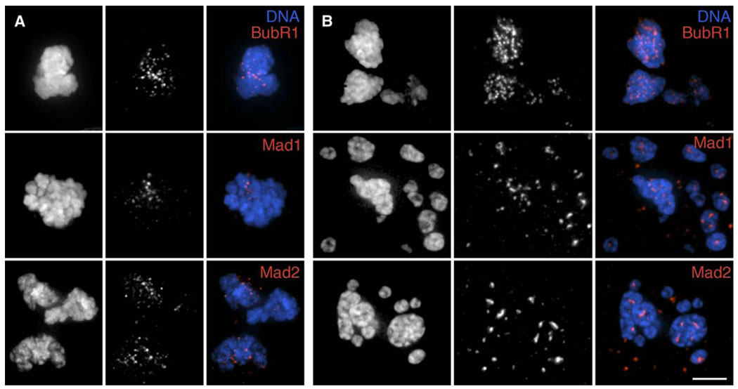
Figure 4. BubR1, Mad1, and Mad2 Remain Associated with RPE1 Kinetochores as Cells Exit Mitosis in the Presence of an Unsatisfied SAC.
RPE1 cultures were incubated with 200 nM (A) or 3.2 µM(B) nocodazole for 9 or 15 hr, respectively, before fixation and immunostaining for BubR1 (top), Mad1 (middle), or Mad2 (bottom). Cells that had just exitedmitosis (see text for details) were then located and photographed. Left columns in each panel, DNA; middle columns, BubR1, Mad1, or Mad2; right columns, merged images. Scale bar equals 5 µm.

