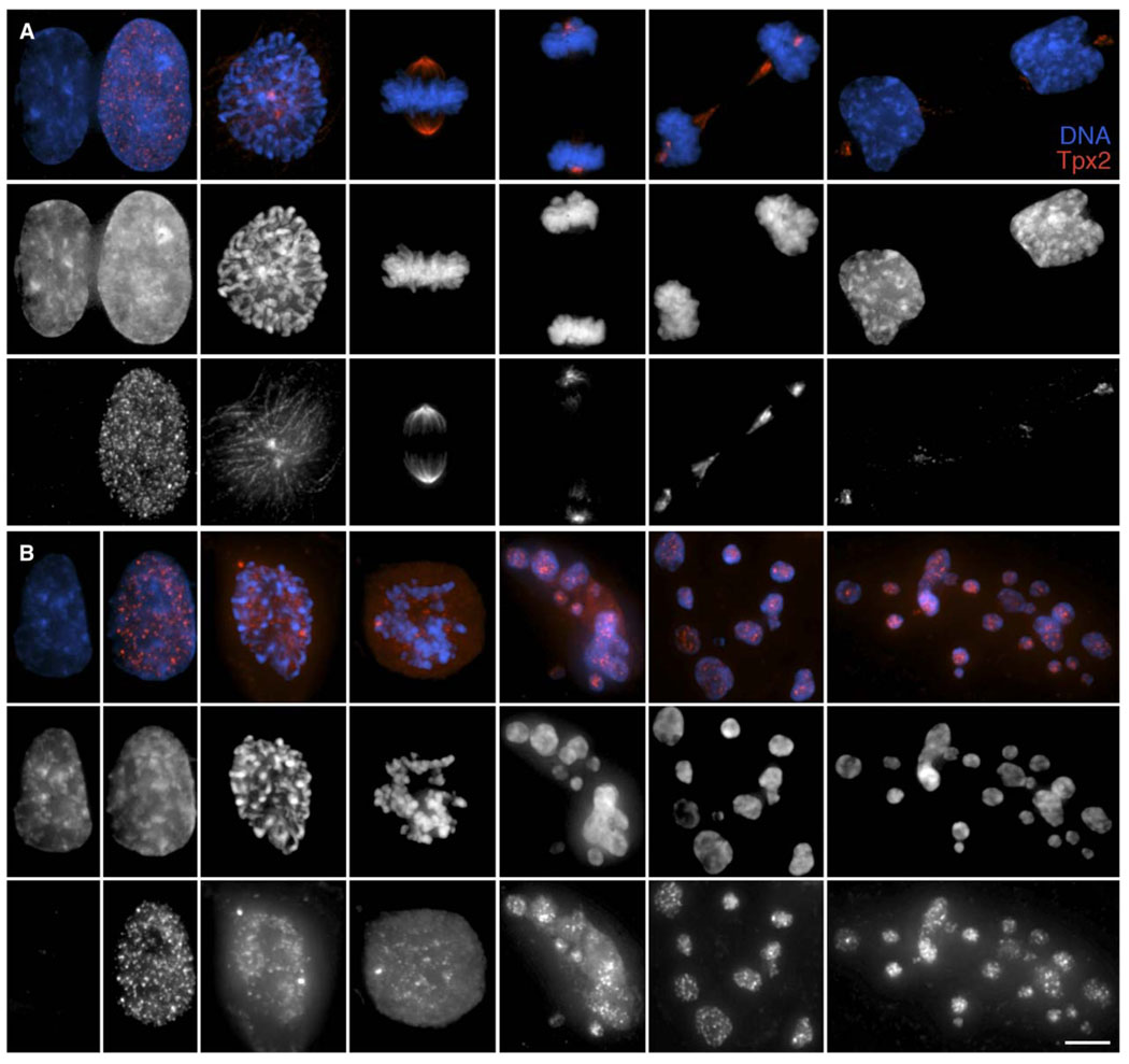
Figure 5. Tpx2, an APC Substrate, Is Not Degraded in Cells that Exit Mitosis without Satisfying the SAC.
(A) In untreated RPE1 cells, Tpx2 (bottom) is found in S/G2 but not G1 nuclei. During mitosis, this MT-associated protein localizes to spindle MTs, while during anaphase it is found on midbody MTs and centrosomes. Tpx2 is largely degraded by the time the nuclear envelope reforms around daughter nuclei during late telophase, although some remains associated with the centrosomes (far right row).
(B) After treatment with 3.2 µM nocodazole, Tpx2 (bottom row) is found in the nuclei of G2 and prophase cells. In the absence of spindle MT formation, this protein then remains diffuse in the cytoplasm throughout the prolonged mitosis. However, once the cells exitmitosis and reflatten on the substrate (last three images in each row), it is found concentrated in the G1 micronuclei. Top row, merged images; middle row, DNA; bottom row, Tpx2. Scale bar equals 5 µm.

