Figure 6.
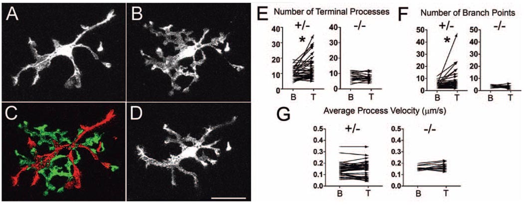
Exogenously applied CX3CL1 leads to changes in microglial morphology. Representative example of a CX3CR1+/− retinal microglial cell (A) under baseline conditions and (B) after application of exogenous CX3CL1 (12.5 nM for 10 minutes). (C) In response to CX3CL1, microglia retract their longer processes (red) and produce multiple highly branched lamellipodia-like processes (green), as shown in the subtraction image. These morphologic changes were reversible, with microglia reverting back to their usual morphology after washout of CX3CL1 (D, 12 minutes after washout). No migratory behavior involving the translocation of the soma was observed during or after CX3CL1 treatment over approximately 30 minutes. (E) Quantification of terminal processes in retinal microglia under baseline conditions (indicated as “B” on the x-axis) and during CX3CL1 treatment (10–15 nM; indicated as “T”). After CX3CL1 application, the number of their processes significantly increased in CX3CR1+/− microglia, whereas little change was seen with CX3CR1−/− microglia. *P < 0.05. (F) The number of branch points similarly increased significantly with CX3CL1 application in CX3CR1+/− microglia, but not in CX3CR1−/− microglia. *P < 0.05. (G) Average process velocity was not significantly affected by CX3CL1 treatment in CX3CR1+/− or CX3CR1−/− retinal microglia.

