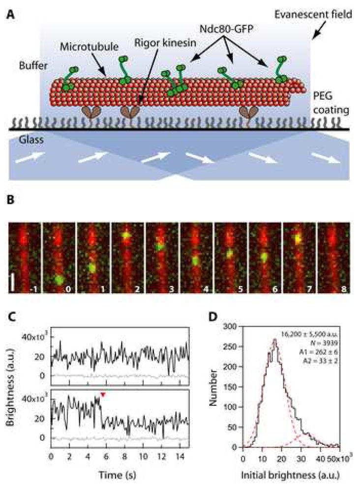
Figure 4. Ndc80 complexes exhibit one-dimensional diffusion along microtubules.

(A) Schematic of the TIRF assay for observing GFP-tagged Ndc80 complexes (green rods) interacting with individual microtubules (red). Excitation by total internal reflection, in combination with a surface treatment that blocks non-specific adsorption, allowed movement of single Ndc80 complexes to be recorded in the evanescent field.
(B) Selected frames from Movie S3 showing one-dimensional diffusion of the Ndc80 complex (green) along a microtubule (red). Elapsed times are in seconds. Scale bar, 1μm.
(C) Records of brightness versus time for most diffusing particles were roughly constant over time (as in the upper trace). However, some particles showed a stepwise loss of half their intensity while they remained attached to the filament (red arrowhead, lower trace), consistent with photobleaching of one GFP molecule within a particle containing two GFPs.
(D) Distribution of initial brightness values for particles of Ndc80-GFP diffusing on taxol-stabilized microtubules. Data are fit by the sum of two Gaussians (dashed red curves), corresponding to a large population (89%) with a unitary brightness of 16,200 ± 5,500 a.u. (mean ± s.d.) plus a small population (11%) with twice the brightness, 32,400 ± 5,500 a.u. (N = 3,939 events on 172 microtubules in 34 recordings).
