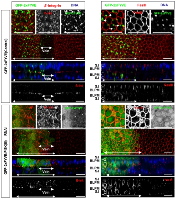
Figure 5. Subcellular localization defects of β-integrin and Fas III in PI3K (III)-knockdown pupal wings.
Confocal immunodetection showing XY and XZ sections in the plane of pupal wings at 30 h APF. Double-headed arrows indicate the regions where the dsRNA for dVps15 was expressed by dpp-Gal4 driver. β-integrin [β-int] (red) was localized to the basal plasma membrane in the wild-type prospective intervein regions but was only rarely localized to the basal plasma membrane while it accumulated in the cytoplasm in dVps15-knockdown cells. β-integrin was not degraded in the knockdown vein region neither was it observed in the wild-type vein region. By contrast, Fas III (red) was localized only to the SJ (indicated by brackets) in the wild-type while in the dVps15-knockdown cells it was accumulated to both the SJ and BLPM regions. The scale bars represent 10 µm.

