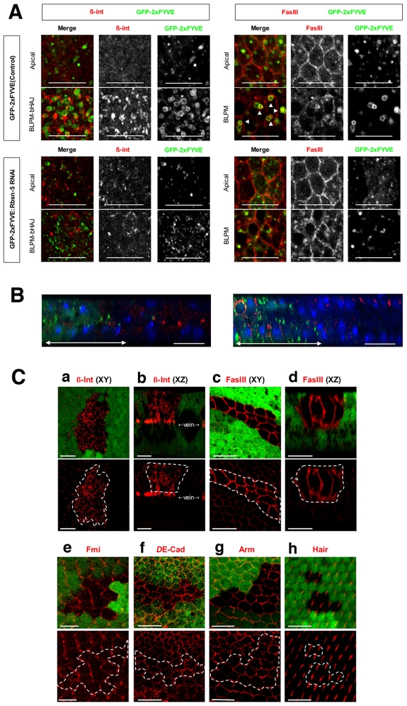
Figure 6. Rbsn-5 was selectively required for the correct localization of FasIII and β-integrin.
(A) Horizontal sections of 27 h APF pupal wings, expressing GFP-2xFYVE, with or without dsRNA, for Rbsn-5 (lower or upper panels, respectively). Images were obtained for repeated 1 µm sections from the apical side. Merged horizontal sections in the plane of the ZA and septate junctions are shown as Apical and those in the plane between the BLPM and bHAJ are indicated as BLPM-bHAJ. β-integrin was mostly localized to the basal plasma membrane and formed large clusters. In Rbsn-5 knockdown cells, β-integrin was rarely localized to the basal plasma membrane and there was only a small amount of accumulation in the apical region. Fas III was localized to the SJ, whereas in Rbsn-5 knockdown cells it also accumulated in the BLPM. In addition, two types of GFP-2xFYVE-positive endosomes were observed as dot-like small structures in the apical regions and vesicle-like large structures in basal regions in the wild type. In Rbsn-5 knockdown cells, very few large endosomes were observed in the basal regions. Note that the large endosomes were very close to the BLPM membrane and frequently contained Fas III. (B) Vertical sections of the pupal wings expressing GFP-2xFYVE (green) with dsRNA for Rbsn-5. (left) β-integrin (red) was not localized to the basal junctions in the knockdown cells where the GFP-2xFYVE is expressed (green). (right) Localization of Fas III (red) extended to the BLPM in the knockdown cells (green). The normal distributions of these proteins were presented in the internal control regions where GFP-2xFYVE was not expressed. (C) Confocal immunodetection showing XY (a, c, e, f, g and h) and XZ (b and d) sections in the plane of the pupal wings at 30–32 hr APF. Cells that are encircled by white lines were GFP-negative and mutant for Rbsn-5C241. β-integrin (red in a and b) accumulated intracellularly, as compared with the wild-type cells surrounding the mutant cells. Fas III (red in c and d) accumulated in the whole basolateral plasma membrane, as compared with the GFP-positive wild-type cells. Fmi (red in e), DE-cadherin (red in f), Arm (red in g) and wing hairs (red in h) were normal. The scale bars represent 10 µm.

