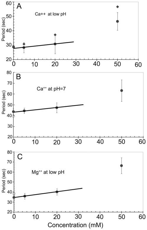
Figure 4. Effect of divalent cations on MinD oscillation periods in E. coli strain PB103.
Fluorescence images of bacteria were recorded 12–15 minutes after introduction of a new ion concentration into the flow cell. (A) Effect of Ca++ ions at an un-buffered pH of 5.5 to 5.8 in the presence of 5 mM NaCl. Raw period data (filled diamonds) have been corrected for cumulative excitation illumination effects (filled circles), as discussed in the text. At 100 mM of Ca++ (data point not shown) bacterial fluorescence was uniform over the cell and no oscillating component was observable. (B) Effect of Ca++ at a pH of 7.0 in 10 mM HEPES buffer (filled circles). The effects of Ca++ cations were reversible, and the original period (open circle) was recovered upon Ca++ removal. (C) Effect of Mg++ ions on the MinD oscillations at low pH in 10 mM HEPES buffer (filled circles). On return to pure buffer the oscillations returned to their initial value (open circle). There is an approximately linear response of the oscillation period to moderate concentrations of extracellular Ca++ or Mg++, as indicated by the solid lines.

