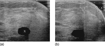
Figure 3.
(a) Slice from prechemotherapy image volume mapped into the space of postchemotherapy image volume. The blacked out region (marked as “X”) was obtained by applying the transformation of the pre- to postchemotherapy registration to the hand-segmented prechemotherapy tumor volume. (b) The corresponding slice in the hand-segmented postchemotherapy image volume for validation.

