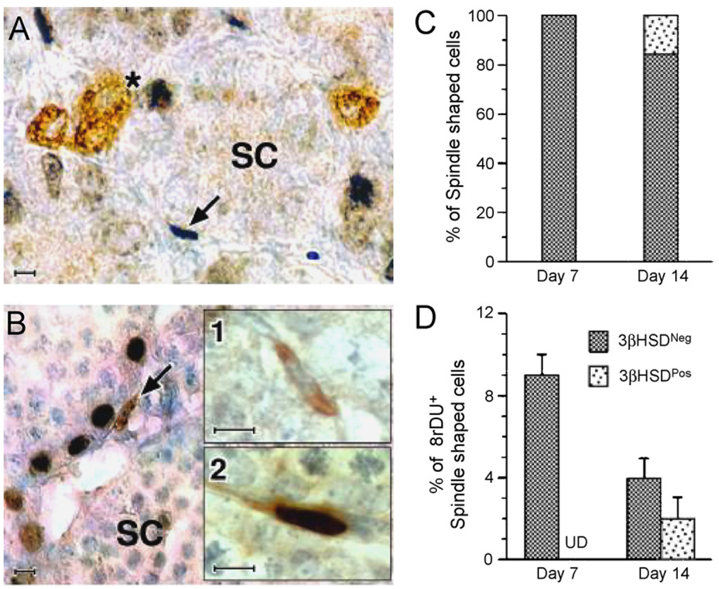
Fig. 2.
Double immunolabeling of testicular cells for 3β-HSD and BrdU in sections of testes from day 7 (A) and day 14(B) rats. A cluster of 3β-HSD-positive cells, presumed to be fetal Leydig cells is immunolabeled (brown staining, indicated by “*”) on day 7 (A). At this age, spindle-shaped interstitial cells, presumed to be stem Leydig cells (SC, indicated by arrow), often were BrdUrd-labeled (dark blue). One week later (day 14, B), spindle-shaped progenitor Leydig cells (brown stained, indicated by arrow) are seen. 3β-HSD-positive spindle-shaped cells were either negative (B1) or positive (B2) for BrdUrd staining. (C and D) The percentages of spindle-shaped cells that were 3β-HSD-positive on days 7 and 14 (C) and BrdUrd-labeled on those days (D). Scale bars, 10 µm. (From Ge et al., 2006 with permission.)

