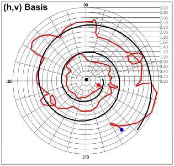
Fig. 6.
Trajectory of CPRs by ex-vivo rodent tail tendon specimen in the (h, v) basis complex plane. The CPR of optic axis (black dot) and noise-free polarization arcs (black trajectory) are estimated from speckle-noise CPRs (red trajectory) (Red and blue dots represents the first and last CPRs, respectively).

