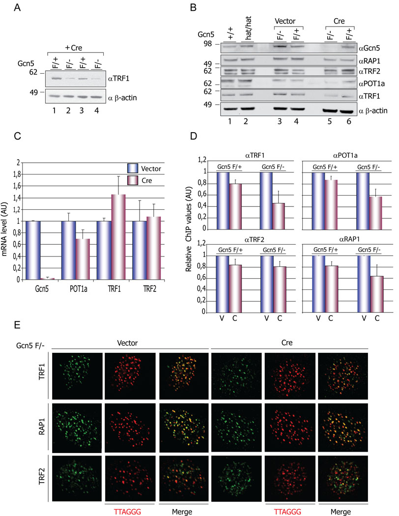
Figure 2. Decreased levels of Shelterin components in Gcn5 null cells.
(A) Decreased levels of TRF1 protein in Gcn5 null MEFs. Protein levels of TRF1 are decreased in Gcn5F/− (lanes 2 and 4) compared to Gcn5F/+ (lanes 1 and 3) MEFs following Cre treatment. (B) Immunoblot data showed decreased TRF1 and POT la protein levels in Gcn5F/− (after Cre treatment) but not in Gcn5F/+ or Gcn5hat/hat cells (lane 5 compared to lanes 1, 2, 3, 4, and 6). Levels of the other shelterin components monitored do not show significant variations in any of the genotypes examined. (C) mRNA level of shelterin components tested by real-time RT-PCR. Experiments were performed with 2 individual MEF cell lines. mRNA level data were normalized by that of GAPDH and vector-treated group values were set as 1. Error bars represent standard deviation from the mean, (D) Decreased amounts on TRF1 and POT la on telomeres. Chromatin immunoprecipitations (ChIPs) were performed using the indicated antibodies and slot blots hybridized with a γ32P-ATP end-labeled TTAGGG probe. The membranes were exposed to Phospholmager screens and the signals quantified with ImageQuant software. Error bars - S.E.M, n=3 independent ChIP experiments. V=vector, C=Cre. (E) Proper localization of TRF1, TRF2 and RAP1 on telomeres. Vector or Cre transfected Gcn5F/− MEFs were immunostained with the indicated antibodies and telomere localization of TRF1, TRF2 and RAP1 was monitored after hybridization with TRITC-conjugated TTAGGG probe.

