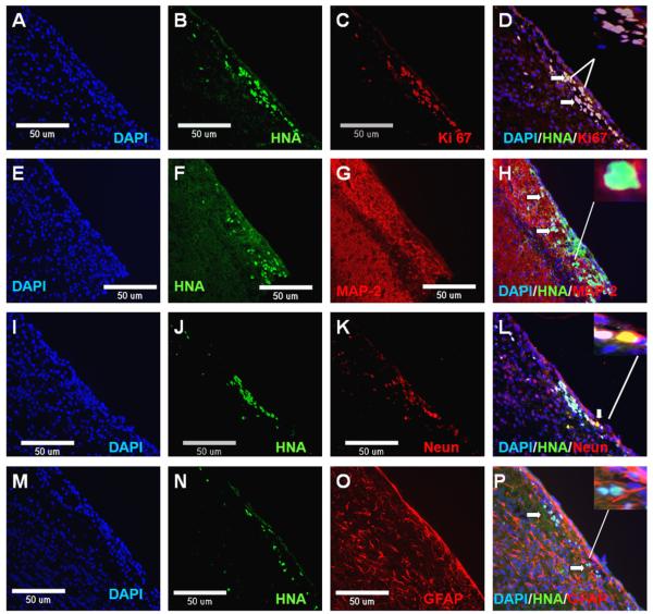
Fig. 7.
Neural rosette cells derived from ORMES6 gave rise to neuron and glial cells in vivo. The rosette cells were transplanted into the striatum of NIHIII mice. At 1, 2, 4 weeks after transplantation, the mice were sacrificed and the brains collected for analysis. The cells could be found at 1 week (n = 3) and 2 weeks (n = 3) post-transplantation. And the number of surviving cells is very small. By 4 weeks, cells were not detected (n = 3). The transplanted cells were distinguished from the host cells by human-specific nuclear antigen (HNA) staining. The transplanted neural rosette cells expressed cell proliferation marker Ki67, astrocyte marker GFAP and neuron markers MAP2 and NeuN. (A) DAPI (blue label); (B) HNA (green label); (C) cell proliferation marker Ki67 (red label); (D) Merge; (E) DAPI (blue label); (F) HNA (green label); (G) neuronal-specific protein MAP (red label); (H) Merge; (I) DAPI (blue label); (J) HNA (green label); (K) neuron marker NeuN (red label); (L) Merge; (M) DAPI (blue label); (N) HNA (green label); (O) astrocyte marker GFAP (red label); and (P) Merge. The nuclei are counter-stained with DAPI (blue). Scale bar: 50 μm.

