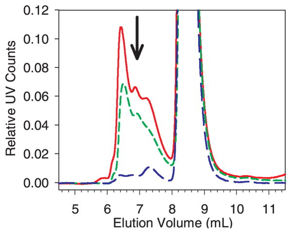
Figure 5.
Zoomed-in view of SEC chromatograms showing the decrease in soluble aggregates when an initially aggregated sample was incubated with cellulose. The y-axis scale of UV counts was normalized to the initial mAb monomer peak height. The arrow shows the trend in peak height/area. Legend: aggregated mAb without added cellulose, solid red line; aggregated mAb after 30 min incubation with cellulose, short-dashed green line; aggregated mAb after 24 hr incubation with cellulose, long-dashed blue line. The color version of this figure can be accessed in the online article.

