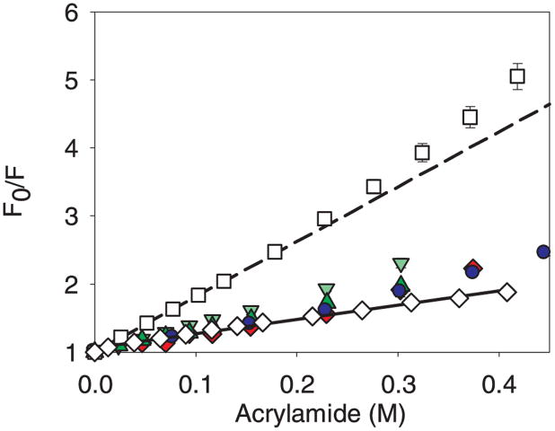
Figure 9.
Stern-Volmer plots for the acrylamide quenching of mAb when free in solution and when bound to microparticles. Legend: native control mAb (◇), with solid line; mAb unfolded in 9 M urea (□), with short dash line; mAb adsorbed to ground glass vials ( ); mAb adsorbed to ground glass syringes (
 ); mAb adsorbed to cellulose (
 ); mAb adsorbed to silica (
 ). Data points are mean ± SD for 3 separate experiments. Error bars may be obscured by data points, and some data symbols overlay. The color version of this figure can be accessed in the online article.

