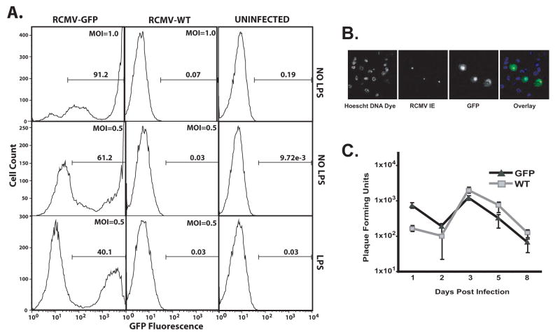
Figure 2. Rat BMDCs Are Highly Susceptible To RCMV Infection.
A) BMDCs were infected 24h post LPS treatment (mDC) or mock (iDC) at an MOI=1.0 (top panel) or 0.5 (middle and bottom panels) for 48h and then assessed by flow cytometry for GFP expression. Gate numbers represent the percent of the population positive for GFP.
B) BMDC were infected with RCMV-GFP at an MOI=0.1. At 48 hours post-infection, the cells were fixed with 1% paraformaldehyde, permeabilized and stained with anti-RCMV IE antibody (red) and with Hoechst (blue), GFP (green). Mag=60x.
C) Growth kinetics of RCMV-WT (squares) and RCMV-GFP (triangles) in BMDCs. At the indicated time points after infection (days post-infection) the presence of virus (combined intracellular and extracellular) in the cultures was determined by standard plaque assays on rat RFL6 fibroblasts. Viral titers are expressed as plaque forming units per ml and represent the average of three replicates.

