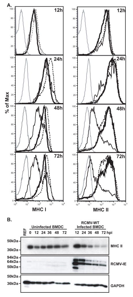
Figure 6. RCMV Induces A Stable Depletion Of Cell Surface MHC I And II.
A. BMDC were infected with RCMV-GFP at an MOI=0.1 and harvested at the times indicated for analysis via FACS. Uninfected cells were harvested in parallel at the indicated times. Isotype control (grey line), Uninfected cells (thick black line), RCMV infected, GFP positive cells (thin black line), BMDC from the same infection culture but uninfected, GFP negative (dashed black line).
B. Western blot analysis of MHC II depletion in RCMV-infected BMDC. BMDC were infected with RCMV-WT at an MOI=1 and the cells were harvested in Laemmli’s sample buffer at the indicated time points after infection. Uninfected rat RFL6 cells and uninfected BMDCs served as controls. Blots were stained for MHC II, RCMV-IE or GAPDH.

