Fig. 5.

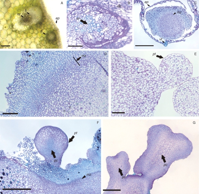
Histological analyses of peach palm somatic embryogenesis. (A) Histological section of fresh tissue to illustrate the organization of a mature tissue evidencing a collateral vascular bundle distributed along the tissue and intercalated with a fibre bundle. (B) First cell division events (arrow) observed in cells adjacent to the vascular tissue and the degenerating parenchyma (dp, arrow). (C) Primary callus induction: note the formation of a meristematic zone (arrowhead). (D) Details from the meristematic zone (arrow). (E) Globular somatic embryo, showing polarization signals and a well-defined protodermis. (F) Globular somatic embryos observed in maturation culture conditions. (G) Mature somatic embryos showing a well-developed procambium and without vascular connection with the matrix tissue. Abbreviations: xy, xylem; fl, phloem; fi, fibre bundle; ep, epidermis; dp, degenerating parenchyma; vt, vascular tissue; pt, protodermis; pc, procambium; mz, meristematic zone. Scale bars: A, B, D, E = 200 µm; C, F, G = 500 µm.
