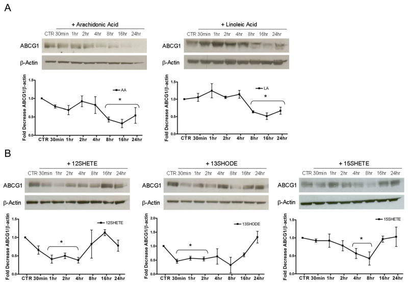
FIGURE 2. 12/15LO activity reduces cellular ABCG1.
Cell lysates were analyzed by immunoblotting for ABCG1 at each time point. Each time point is compared to control (CTR, e.g. time 0) for statistical evaluation. Panel A. Top, J774 macrophages were incubated with vehicle control (CTR) or with arachidonic (AA) or linoleic (LA) acid (125 μM) for up to 24 hours. Bottom, Densitometry of immunoblot normalized to beta actin. Data represent the mean ± SE of 3 experiments (*significantly lower than CTR p<0.04 by ANOVA). Panel B. Top, J774 macrophages were incubated with vehicle control (CTR) or with the indicated eicosanoids (500nM) for up to 24 hours. Bottom, Densitometry of immunoblot normalized to beta actin. Data represent the mean ± SE of 3 experiments (*significantly lower than CTR p<0.05 by ANOVA).

