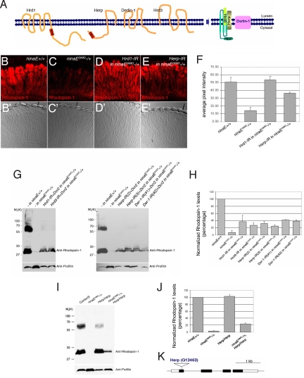
Fig. 1.
Loss of ERAD regulators partially restores the level of Rh-1 in the retina of ninaEG69D−/+ flies. (A) Schematic diagrams of the predicted membrane topology and domain structure of ERAD components through EMBnet-CH. (B–E) Horizontal retina cryosections stained with anti-Rh-1 antibody in 1- to 3-day-old flies. (B) A control ninaE +/+ retina. ninaEG69D−/+ retina (C) show low Rh-1 labeling, while Rh-1 levels recover when Hrd1 (D) or Herp (E) are knocked down in this genetic background. In all RNAi experiments, dicer2 (drc2) were co-expressed to enhance the efficiency of RNAi knockdown. (F) The average intensity of anti-Rh-1 labeling (n = 3), as quantified through the Image J. Error bars, ± SEM. (G and I) A Western blot of Drosophila adult head extracts of the indicated genotypes probed for Rh-1 (upper gel) and anti-profilin (as a loading control; lower gel). (H and J) Quantification of the average normalized Rh-1 band intensity as shown in (G and I) (average of n = 3; Der-1, average of n = 2), with the value from wild-type of Rh-1+/+ heads extracts set at 100%. (K) The structure of the Herp genomic locus. In the HerpG13463, an EP-element is inserted within its protein-coding region. Genotypes: Rh1-Gal4;;ninaE+/+ (B and B′), Rh1-Gal4;;ninaEG69D/+ (C and C′), Rh1-Gal4;UAS-Dcr2/+;UAS-Hrd1-IR/ninaEG69D (D and D′), Rh1-Gal4;UAS-Dcr2/+;UAS-Herp-IR/ninaEG69D (E and E′), (G, lanes 1 and 5) Rh1-Gal4;;ninaE+/+, (G, lanes 2 and 6) Rh1-Gal4;;ninaEG69D/+, (G, lane 3) Rh1-Gal4;UAS-Dcr2/+;UAS-Hrd1-IR/ninaEG69D, (G, lane 4) Rh1-Gal4;UAS-Hrd3-IR/+;UAS-Dcr2/ninaEG69D, (G, lane 7) Rh1-Gal4;UAS-Herp-IR/+;UAS-Dcr2/ninaEG69D, (G, lane 8) Rh1-Gal4;UAS-Dcr2/+;UAS-Herp-IR/ninaEG69D, (G, lane 9) Rh1-Gal4;UAS-Dcr2/+;UAS-Der-1-IR(#1)/ninaEG69D, (G, lane 10) Rh1-Gal4;UAS-Dcr2/+;UAS-Der-1-IR(#2)/ninaEG69D, (I, lane 1) CantonS, (I, lane 2) ninaEG69D/+, (I, lane 3) HerpG13463/HerpG13463, (I, lane 4) HerpG13463/HerpG13463;ninaEG69D/+.

