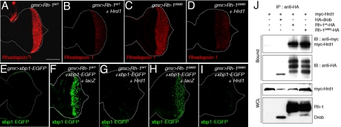
Fig. 2.
ER stress caused by Rh-1 misexpression is strongly suppressed by Drosophila Hrd1. (A–D) Representative images of eye imaginal discs expressing Rh-1WT (A) or Rh-1G69D (C) alone, or together with Hrd1 (B and D). Anti-Rh-1 antibody labeling is in red. E–I Hrd1 co-expression abolished ER stress caused by wild-type or mutant Rh-1 misexpression, as determined by the ER stress marker, xbp1-EGFP (green). Shown are representative discs expressing xbp1-EGFP alone (E), or together with indicated genes. (J) Co-immunoprecipitation assays between Hrd1 and Rh-1 in 293T cells. Hrd1 was tagged with the myc epitope, while Rh-1 was tagged with HA. HA-Drob-1 is a membrane protein used as a negative control. [Scale bars, 100 μm (A).] Genotypes: gmr-Gal4/+;UAS-Rh-1WT/+ (A), gmr-Gal4/UAS-Hrd1;UAS-Rh-1WT/+ (B), gmr-Gal4/+;UAS-Rh-1G69D/+ (C), gmr-Gal4/UAS-Hrd1;UAS-Rh-1G69D/+ (D), gmr-Gal4/+;UAS-xbp1-EGFP/+ (E), gmr-Gal4/UAS-lacZ;UAS-Rh-1WT/UAS-xbp1-EGFP (F), gmr-Gal4/UAS-Hrd1;UAS-Rh-1WT/UAS-xbp1-EGFP (G), gmr-Gal4/UAS-lacZ;UAS- Rh-1G69D/UAS-xbp1-EGFP (H), and gmr-Gal4/UAS-Hrd1;UAS-Rh-1G69D/UAS-xbp1-EGFP (I).

