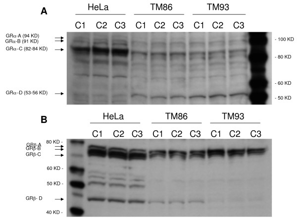
Figure 1.
Western blot analysis of the GRα (A) and GRβ (B) isoforms in human TM86 and TM93. Blots resolving 45 μg of total cell lysates from HeLa, TM 86, or TM 93 cells detected with anti-GRα antibody (PA1-516) or anti-GRβ antibody (PA3-514) are shown. Three independent cell lysates were analyzed per cell line. Additional bands in between GR-C and GR-D isoforms may be degradation products or cell-specific isoforms.

