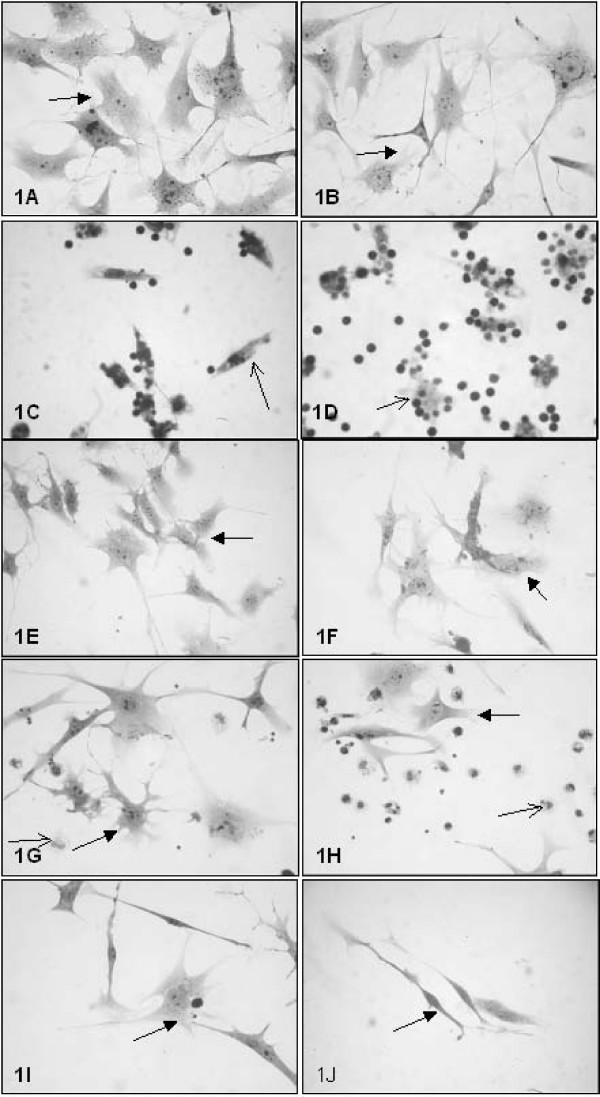
Figure 1.
Microphotographs of control and treated cultures. The cells were fixed, stained with Giemsa, and observed under a light microscope. Left column: control culture conditions; right column: treated culture conditions. (A and B) B16F10 cells; (C and D) Mϕ/Ly; (E and F) B16F10/Ly; (G and H) B16F10/Mϕ; (I and J) B16F10/Ly-Mϕ. Original magnification: 40× objective for all figures. Black arrow: B16F10 cells; thin black arrow: macrophages.

