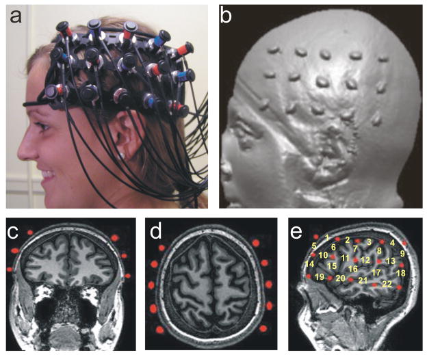
Figure 1.
Hitachi ETG-4000 Imaging System, Neuroanatomical Probe Positioning, and MRI Neuroanatomical Co-Registration. (a) Participant with Hitachi 24-channel ETG-4000, with lasers set to 698nm and 830nm, in place and ready for data acquisition. The 3×5 optode arrays were positioned on participants’ heads using rigorous anatomical localization measures including 10 × 20 system and MRI coregistration (see b-e). (b) MRI co-registration was conducted by having participants (N=9) wear 2 3×5 arrays with vitamin-E capsules in MRI. (c-e) anatomical MRI images were analyzed in coronal (c), axial (d) and sagital (e) views allowing us to identify the location of optodes (Vitamine E capsules) with respect to underlying brain structures. (e) Anatomical view of the position of the fNRIRs channels.

