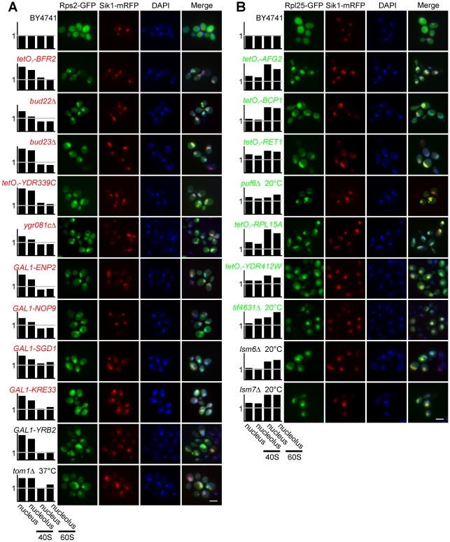
Figure 7. Identification of ribosomal subunit nucleolar and nuclear export defects.
(A) Mutants with observed small subunit-export defects. (B) Mutants with large subunit-export defects. The first row of panel (A) and panel (B) represent the wild-type strain BY4741 cultured at 30°C. Rps2-GFP and Rpl25-GFP are reporters for the ribosomal small and large subunits, respectively. Sik1-mRFP is the reporter for the nucleolus. DAPI was used to stain DNA to visualize the nucleus. The white scale bar represents 5 µm. The normalized enrichment of ribosomal subunits in the nucleus or nucleolus relative to the cytoplasm, calculated relative to the appropriate control strain, is plotted for each strain.

