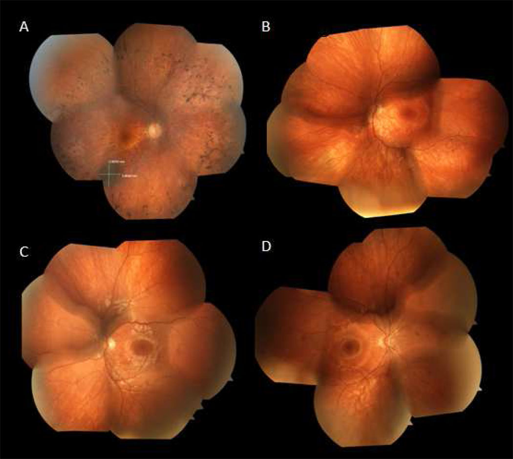
Figure 1. Color fundus photographs.
A. 47 year old man with D190N adRP, right eye. Optic nerve has mild pallor and there is extensive intraretinal pigment migration in the mid-periphery. Note the incidental choroidal nevus 3.9 × 3.36 mm along the inferotemporal arcade.
B. 11 year old son affected with adRP, left eye. Note the mild RPE migration inferonasally.
C. 9 year old son without RP, left eye. Optic nerve without pallor and macula with good foveal reflex.
D. 7 year old son affected with adRP, right eye. Optic nerve without pallor and macula with good foveal reflex, clinically indistinguishable from panel C.

