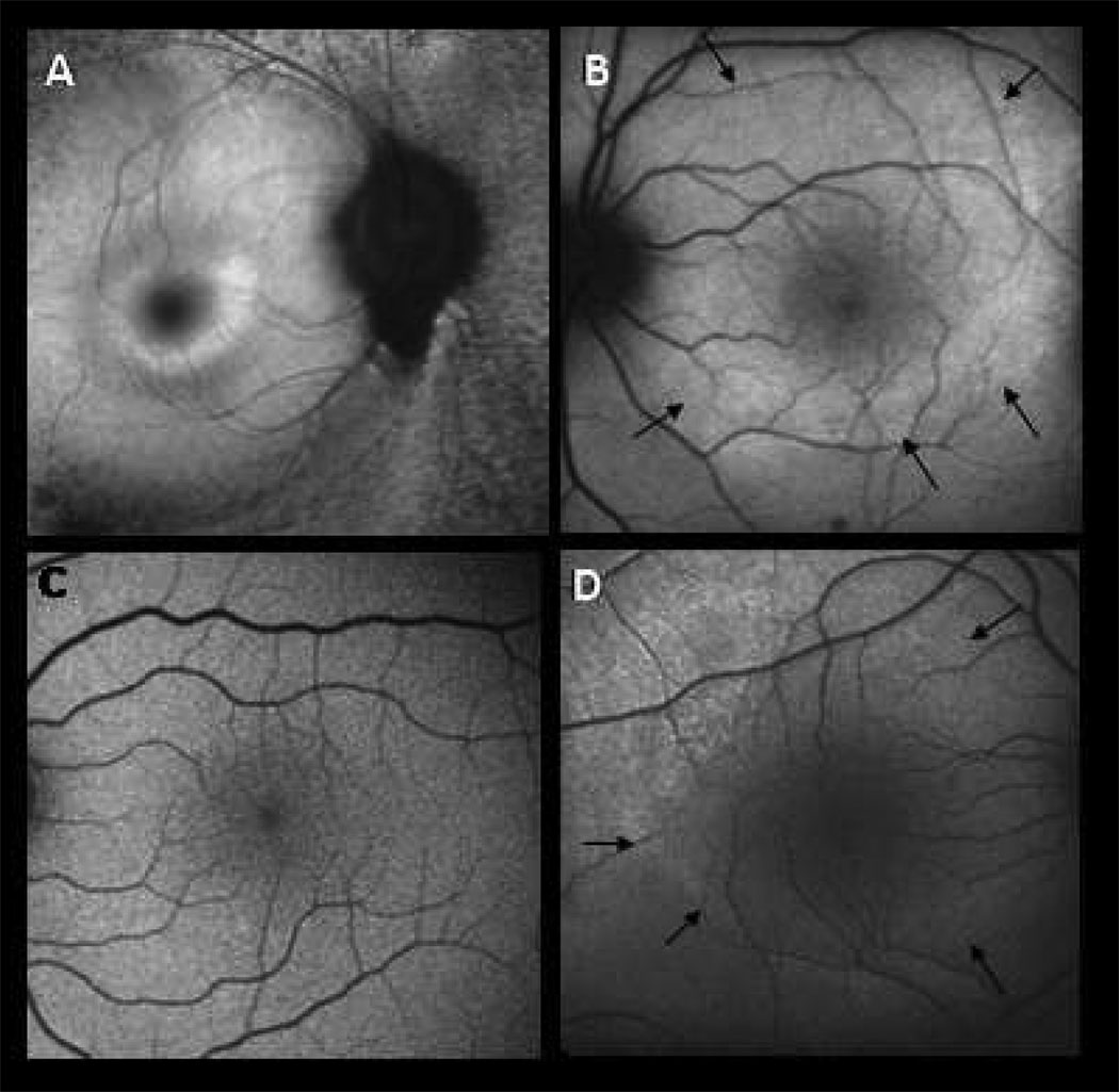
Figure 2. Autofluorescence imaging.
A. 47 year old man with D190N adRP, right eye. Macula with small hyperfluorescent ring.
B. 11 year old son affected with adRP, left eye. Macula with large hyperfluorescent ring (black arrows.)
C. 9 year old son without RP, left eye. Macular autofluorescence normal.
D. 7 year old son affected with adRP, right eye. Macula with early, large hyperfluorescent ring (black arrows) similar to panel B.

