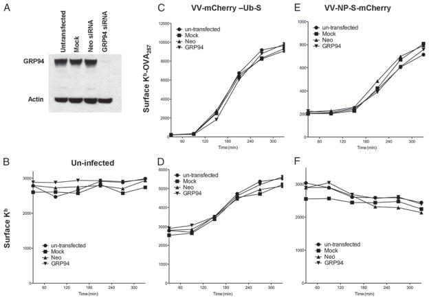
FIGURE 1.
GRP94 has no essential role in Ag processing and presentation. A, Knockdown of GRP94 was confirmed by Western blot analysis. L-Kb cells were untransfected (●), mock transfected (■), transfected with control Neo siRNA (▲), or transfected with GRP94 siRNA (▼). Cells were infected 4–5 days later with VV-mCherry-Ub- S (C and D) or VV-NP-S-mCherry (E and F) or uninfected as a control (B). Samples were taken every 1h up to 5.5 h after infection. Levels of surface Kb-OVA257 were then measured by staining with Alexa Fluor 647-labeled 25-D.1.16 (C and E), and levels of the surface Kb molecule (B, D, and F) were determined by staining with anti-Kb Ab (HB176) followed by a secondary FITC-conjugated anti-mouse Ab. The experiment was repeated two times.

