
Figure 6. Phospho-p38 MAP kinase increases in outer hair cells with aging.
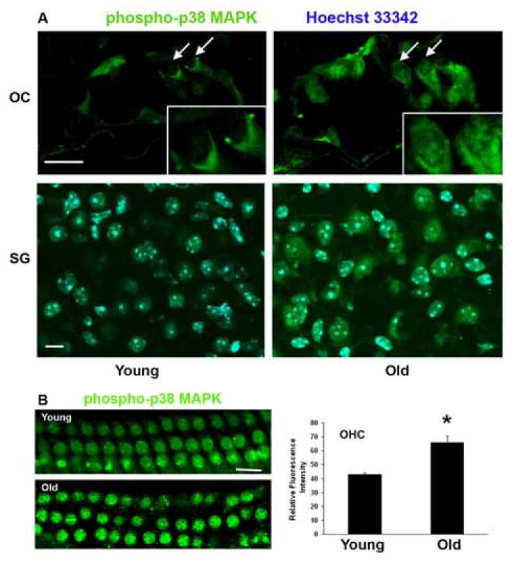
A: Fluorescent staining for phospho-p38 MAPK increased in the organ of Corti with aging. Staining was localized to the nuclei and cytoplasm of outer hair cells of the basal turn (arrows; boxed areas are high magnifications of outer hair cells) and to spiral ganglion cells. Green, phospho-p38 MAPK; blue (in the SG preparation), Hoechst 33342 for nuclei. The figure is representative of 4 individual preparations at each age. Scale bars = 10 μm.
B: Surface preparations of the cochlear epithelium of the basal turn were stained with phospho-p38 MAPK (green) and showed an increase in phospho-p38 MAPK in outer hair cells with age. Quantitative analysis of relative fluorescence intensity confirmed its elevation in outer hair cells. *p < 0.01, n = 3. Scale bar = 10 μm.