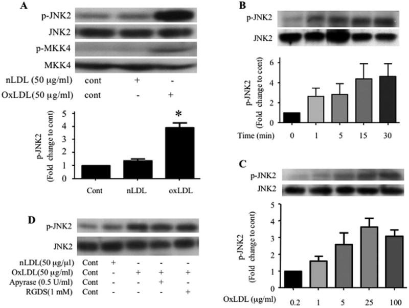
Figure 1. OxLDL induces phosphorylation of JNK2 and MKK4 in platelets.
Washed human platelets (2 × 10⁸/ml) containing 2 mM CaCl2 and 1 mM MgCl2 were incubated with native LDL or various concentrations of oxLDL over varying time points and then lysed. The lysates were analyzed by immunoblot with antibodies specific for phospho–JNK (p-JNK2, A, B, C), phospho-MKK4 (p-MKK4, A). The membranes were then stripped and re-probed with antibodies to the total relevant proteins to normalize the protein loaded. (D) Platelets were incubated with 50 μg/ml native LDL (Lane 2) or oxLDL with PBS (Lane 3), 0.5 U/ml apyrase (Lane 4) or 1 mM RGDS (Lane 5) for 5 minutes and then lysed. The lysates were analyzed by immunoblot as above for phospho-JNK (p-JNK) and total JNK. Results are representative of at least 3 independent experiments from different donors. The bar graph represents quantification of the phosphorylation of JNK2 (ratio of phosphorylated/total) expressed as relative values when compared with platelets without any treatment (A, B) or platelets treated with 0.2 μg/ml oxLDL (C). n=5 for A, n=3 for B and n=4 for C. c- control, nLDL- native LDL, oxLDL-oxidized LDL, * p < 0.05 when compared to control or nLDL treatment.

