
Figure 4.
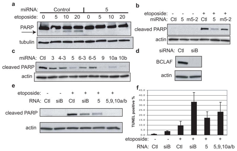
Apoptosis assays in HUVEC with various miRNAs. a, Cells were transfected with miRNAs (control or miR-K5) for 48 hr., followed by 24 hr. treatment with etoposide (μM conc.). Samples were probed with PARP antibody (recognizes full length and cleaved form) and the loading control antibody, tubulin. Arrow indicates cleaved PARP. b, Similar to (a) except cells were transfected with miRNAs (negative control “Ctl”, miR-K5 “5”, or mutant miR-Km5-2 “m5-2”) for 24 hr., followed by 24 hr. treatment with 50 μM etoposide. In this case, samples were probed with antibodies specific for the cleaved PARP chain, and for the loading control, actin. c, All cells shown were transfected with miRNAs for 48 hr., followed by 24 hr. treatment with 20 μM etoposide. Samples were probed with antibodies specific for cleaved PARP and for the loading control, actin. d, Western blot analysis of endogenous BCLAF1 and loading control, actin, in HUVEC cells transfected with siRNAs directed to BCLAF1 (siB) or control (ctl). e and f, HUVEC cells were transfected with the indicated miRNAs or siRNAs, split to low density and treated with 50 μM etoposide followed by western blotting (e) TUNEL staining and flow cytometry (f). Values reflect average from at least three experiments with errors bars showing 1 std. dev.