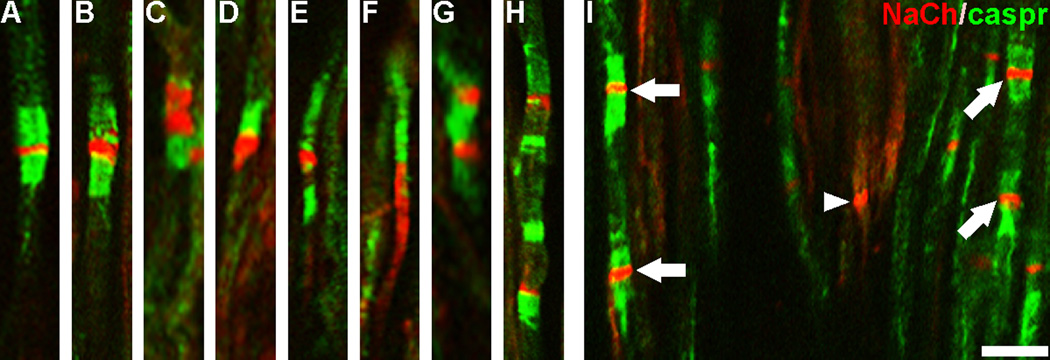
Figure 5.
NaCh and caspr relationships at nodal sites are altered in painful dental pulp. Confocal micrographs of individual image slices show NaCh (red) and caspr (green) staining relationships in a normal (A) and painful (B–I) samples. The NaChs are located in the narrow nodal gap and are flanked by dense paranodal caspr staining in a typical node from a normal sample (A), whereas deviations from this relationship are seen in NaCh accumulations identified in painful samples. These deviations include enlarged NaCh accumulations that show a decreased intensity or even an elimination of caspr staining on one side of the NaCh accumulation (B–F), while other single fibers show multiple NaCh accumulations (arrows) separated by short distances that show atypical (G–H) and typical (I) caspr relationships. Some NaCh accumulations lacked associations with caspr (arrowhead). Scale bar = 10 µm.

