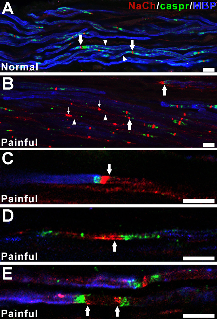
Figure 6.
Painful samples show NaCh expression in fibers with decreased staining for myelin basic protein (MBP). Confocal micrographs of collapsed z-projection images (A, B; five separate image slices separated by 1 µm increments) and single image slices (C–E) show NaCh (red), caspr (green) and MBP (blue) staining relationships within a normal sample (A) and variations from this pattern in painful samples (B–E). Myelinated fibers within the normal dental pulp (A) show prominent surface staining for MBP (arrowheads) and NaCh accumulations at caspr-identified typical nodal sites (arrows). In contrast, the painful sample (B) shows generalized and focal areas of decreased MBP staining (arrowheads) and prominent NaCh accumulations at sites that lack caspr (small arrows) and at other sites that show alterations in caspr relationships (large arrows). The remodeling of NaChs at caspr-identified atypical nodal sites within single fibers that show decreased staining for MBP is shown in C–E. This remodeling includes prominent NaCh expression within axon segments that are flanked by caspr and that lack MBP staining (C–E; arrows). These findings are consistent with the remodeling of NaChs at demyelinated sites within the painful dental pulp. Scale bars = 10µm.

