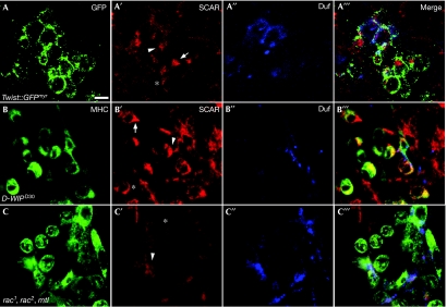
Figure 2.
Localization pattern of SCAR in migrating and fusing myoblasts. (A–A′″) A stage 13 wild-type twist-GAL4∷UAS–GFPmyr embryo stained with anti-GFP (A, green) to visualize the contours of myoblasts, anti-SCAR (A′, red) and anti-Duf (A′′, blue), which mark myotube surfaces. The asterisk marks a rounded (pre-migratory) myoblast in which SCAR is uniformly localized in the membrane; an arrow points to a ‘tear'-shaped, migrating myoblast, in which SCAR localizes to the lamellipodium; and the arrowhead to a myoblast–myotube fusion interface. (B–B′″) A stage 14-D-WIPD30 embryo. Anti-MHC (B, green) was used as the general myoblast marker. SCAR localization within the enriched population of unfused myoblasts resembles that seen in wild-type embryos. Cells are marked as in A′,A′″. (C–C′″) A rac triple-mutant embryo stained as in (B–B′″). SCAR is uniformly distributed within the unfused myoblasts, all of which show a rounded morphology. Scale bar, 10 μm. GFP, green fluorescent protein; MHC, myosin heavy chain; SCAR, suppressor of cyclic AMP receptor; UAS, upstream activation sequence.

