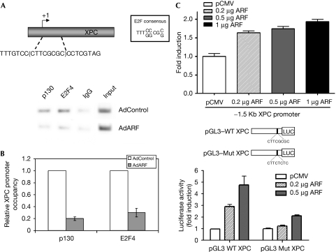
Figure 5.
ARF stimulates the expression of XPC by disrupting the repressor complex of E2F4. TKO MEFs were infected with either control or ARF-expressing adenovirus and subjected to ChIP assay. XPC promoter DNA associated with immunoprecipitated chromatin was quantified by using real-time PCR. An aliquot of the RT–PCR sample was analysed by agarose gel electrophoresis (A), and the quantitative RT–PCR results are shown in panel (B). In panel (C), NIH-3T3 cells were serum starved for 12 h followed by transfection with a 1.5 kB fragment of the XPC and a smaller fragment of the promoter (−51 to +7) containing the E2F site or a mutant promoter construct along with the indicated amounts of ARF. Luciferase activity was measured. Results from three experiments were plotted. ARF, alternative reading frame; ChIP, chromatin immunoprecipitation; E2F4, E2F transcription factor 4; MEF, mouse embryonic fibroblast; Mut, mutant; RT–PCR, reverse transcription PCR; TKO, triple knockout; WT, wild type; XPC, xeroderma pigmentosum group C.

