Figure 5.
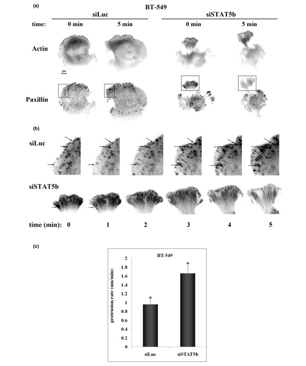
STAT5b-knockdown cells exhibit multiple protrusions with increased retraction. BT-549 cells were transfected with siRNA (siLuc or siSTAT5b), GFP-speckle-actin, and mKO-paxillin. Seventy-two hours after transfection, cells were plated on 3 μg/ml fibronection (FN) for 20 to 30 minutes, and TIRF time-lapse microscopy was performed. Images were taken every 3 seconds for 5 minutes at 60× magnification. (Line indicates 10 μm). (a) Still images representing actin and paxillin staining in control (siLuc) and STAT5b-knockdown (siSTAT5b) cells at start of filming (0 min) and end of filming (5 min). (b) Enlarged images depicting boxed areas from part a. Arrows identify single focal adhesions at each time point. (c) Protrusion rate (μm/min) was calculated as the length of the protrusion divided by the total time of the movie for at least three independent experiments (siLuc, n = 16; siSTAT5b, n = 13). Student's t test was used to determine statistical significance (P < 0.05) between siLuc and siSTAT5b (*).

