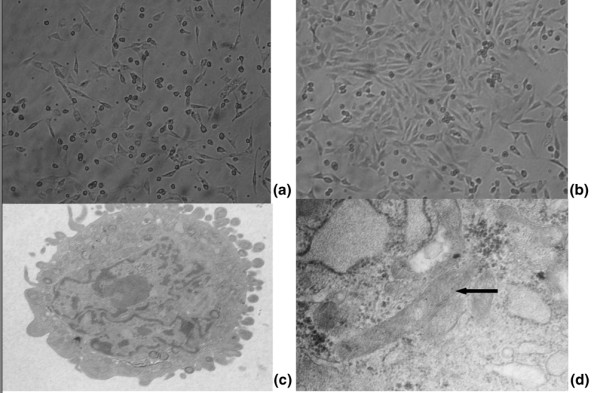
Figure 2.

Endothelial progenitor cell morphology during ex vivo culture. (a) Mononuclear cells are able to differentiate into spindle cells 48 hours after culture.(b) Colony of endothelial progenitor cells (EPCs) observed after 7 or 8 days of culture. (c) and (d) After 7 days of culture, the ultrastructure of EPCs can be observed by electron microscope. Black arrow, Weible-Palade body that was the characteristic structure of EPCs. Magnification: (a) and (b) ×100; (c) ×5,000; (d) ×30,000.
