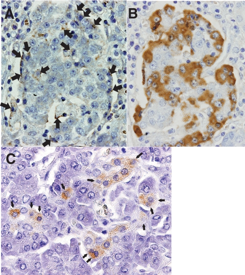
FIG. 1.
A: Immunohistochemical demonstration of enterovirus-associated VP1 antigen in pancreatic islets (brown, arrows). Cells with shrunken and dark nuclei (arrows) suggestive of pyknosis, a sign of cell death, were observed (×400, case 1). B: Immunohistochemical staining for glucagon in serial sections of (A) (×400). Comparing (A) and (B) indicates enterovirus VP1 antigen residing on islet cells. C: Homogeneous staining for VP1 was observed in pancreatic acinar cell clusters (brown) with shrunken and darkly staining nuclei suggestive of pyknosis (arrows) (×400). (A high-quality digital representation of this figure is available in the online issue.)

