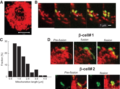
FIG. 1.
Dissection of the mitochondrial web in primary β-cells using PAGFPmt. A: Projection of confocal images of a cell stained with TMRE. Note that mitochondria are densely packed in β-cells. B: Sequential 2-photon laser photoactivation of individual mitochondria. C: Summary of mitochondrial size distribution performed by 90 photoactivation steps in 9 cells. D: Tagging and tracking individual mitochondria in primary β-cells reveals fusion events (transfer of activated PAGFPmt to juxtaposed unlabeled mitochondria) that are then followed by fission events (separation of previously connected mitochondria). (A high-quality digital representation of this figure is available in the online issue.)

